Laser is recommended when a specific eye problem can be stabilised, sealed, or treated with less risk than observation, injections, or surgery. By the end, you will know which retinal findings need urgent assessment, what each laser procedure is designed to do, and what questions to ask before treatment.
Key takeaways
- Seek same-day retinal assessment for flashes, sudden floaters, a curtain, or sudden vision loss.
- Retinal laser seals selected tears and treats abnormal blood vessels; it does not repair every detachment.
- Dilated examination, OCT, retinal photographs, and fluorescein angiography guide treatment decisions.
- Glaucoma laser, YAG capsulotomy, and LASIK treat different eye structures and problems.
Which eye symptoms need urgent retinal assessment?
Arrange a same-day dilated retinal examination for new flashes in vision, a sudden increase in floaters, a curtain or shadow in vision, or sudden vision loss. These symptoms can signal a retinal tear, retinal detachment, bleeding, or another sight-threatening problem that cannot wait for a routine laser consultation.
| Symptom | Why it matters | What to do |
|---|---|---|
| New flashes in vision | Traction on the retina can create a tear | Seek same-day retinal assessment, especially with floaters |
| Sudden increase in floaters | New dark spots, strands, or a shower of dots can indicate bleeding or a tear | Use an emergency eye service today |
| Curtain or shadow in vision | A spreading field defect is a warning sign for retinal detachment | Go immediately; do not wait for symptoms to settle |
| Sudden vision loss | Retinal vascular blockage, detachment, bleeding, or optic-nerve disease can cause permanent damage | Seek emergency assessment now |
| Painful red eye with blurred vision | A pressure spike or severe inflammation can threaten vision | Obtain urgent eye care |
A retinal tear is not the same as an established retinal detachment. Laser retinopexy can seal selected high-risk tears, but laser cannot reattach a detached retina or restore photoreceptors that have died; surgery such as vitrectomy, scleral buckle, or pneumatic retinopexy may be necessary.
Avoid driving yourself if vision is impaired, and take the exact time symptoms began.
When is retinal laser recommended for specific conditions?
The choice depends on what threatens sight: a break that could lead to detachment, abnormal vessel growth, or leakage in the macula. Laser is not automatically the best treatment; injections, observation, cryotherapy, or retinal surgery may carry a better risk-benefit balance.
| Condition | Usual approach | When it applies |
|---|---|---|
| Retinal tears | Laser retinopexy | A symptomatic horseshoe tear or another high-risk tear is surrounded by burns to form a scar barrier. An established detachment needs vitrectomy, scleral buckle, or pneumatic retinopexy instead. |
| Retinal holes | Observation or laser retinopexy | Small, asymptomatic atrophic holes often need monitoring. Laser becomes more compelling when symptoms, subretinal fluid, a detachment in the other eye, or other high-risk findings are present. |
| Lattice degeneration with tear risk | Observation or preventive laser | Lattice alone is not routinely lasered. Treatment is considered when a related tear, symptoms, or substantial detachment risk makes a scar barrier worthwhile. |
| Proliferative diabetic retinopathy | Panretinal photocoagulation, anti-VEGF injections, or both | PRP treats peripheral retina driving abnormal vessel growth. It can reduce severe visual loss but may reduce peripheral or night vision and worsen macular edema. |
| RVO-related neovascularisation | PRP; injections for macular edema | PRP treats new vessels caused by retinal vein occlusion. Anti-VEGF injections usually address fluid and swelling. |
| Selected macular leakage | Focal or grid laser, often after assessment | Focal burns target leaking points; grid treatment covers diffuse leakage. Center-involving diabetic macular edema often receives anti-VEGF injections first. |
Laser creates permanent retinal burns, so the untreated disease must pose a greater danger than the damage treatment causes. Retinopathy of prematurity is a separate, time-sensitive specialist case: peripheral avascular retina may be lasered, with anti-VEGF treatment used for selected aggressive disease.
What tests determine whether retinal laser is appropriate?
A dilated retinal examination often makes the first treatment decision: it shows whether a tear is high-risk, whether the retina is detached, and whether abnormal new vessels are present. Imaging adds detail and a record of change.
| Test | What it shows | How it guides treatment |
|---|---|---|
| Dilated retinal examination | Tears, holes, detachment, bleeding, new vessels | Laser retinopexy for a high-risk tear; vitrectomy or another operation for detachment |
| Optical coherence tomography | Cross-sectional retinal swelling, fluid, or traction | Injections, observation, or selected macular laser instead of broad laser |
| Fundus photography | A documented view of lesions and their progression | Confirms treatment targets and supports follow-up comparisons |
| Fluorescein angiography | Leaking vessels, blocked circulation, and non-perfused retina | Focal or panretinal laser, injections, or observation according to the leakage pattern |
| Visual-field testing | Functional loss, including peripheral defects | Shows whether disease or treatment threatens useful vision |
| Intraocular-pressure measurement | Eye pressure and glaucoma risk | May direct pressure-lowering medication or glaucoma treatment, not retinal photocoagulation |
The findings usually lead to one of these choices:
- Laser seals a threatening tear, treats selected leakage, or ablates ischemic retina driving new vessels.
- Anti-VEGF injections usually take priority for significant macular edema from diabetic disease or retinal vein occlusion.
- Observation suits a stable lesion whose treatment risk exceeds its immediate danger.
- Cryotherapy treats a peripheral tear when laser access is poor.
- Medication controls diabetes, hypertension, inflammation, or eye pressure but does not reattach a detached retina.
- Vitrectomy is considered for established detachment, traction, or persistent vitreous haemorrhage.
How are glaucoma lasers, YAG treatment, and LASIK different?
They differ by the tissue treated, the problem targeted, and the vision change you can realistically expect.
| Procedure | Target and purpose | Expected benefit and limit |
|---|---|---|
| Retinal photocoagulation | Retinal laser burns seal leaking vessels or create a scar barrier around threatened retina. | Controls complications such as abnormal vessel growth or selected leakage; it does not restore damaged retina and can leave permanent blind spots. |
| Selective laser trabeculoplasty | Treats the drainage angle in open-angle glaucoma to improve fluid outflow. | Lowers eye pressure and may reduce reliance on drops; it cannot reverse established optic-nerve damage. |
| Laser peripheral iridotomy | Creates a small opening in the iris when the drainage angle is narrow or at risk of closing. | Reduces the risk of an angle-closure attack; it does not treat glaucoma damage already present. |
| YAG capsulotomy | Clears posterior capsule opacification, a hazy membrane behind an artificial lens after cataract surgery. | Often sharpens vision quickly, but it neither removes nor replaces the lens and will not correct macular, corneal, or glaucoma-related loss. |
| Refractive laser surgery | LASIK or PRK reshapes the cornea to correct short-sight, long-sight, or astigmatism. | Reduces glasses dependence; it does not treat retinal disease, glaucoma, cataract, or optic-nerve damage. |
A retinal laser treats disease, whereas LASIK changes focusing power. SLT and laser peripheral iridotomy protect pressure control or drainage; YAG capsulotomy addresses postoperative haze. Each requires a different examination before treatment, and none is interchangeable.
What happens during laser treatment, and when is another option safer?
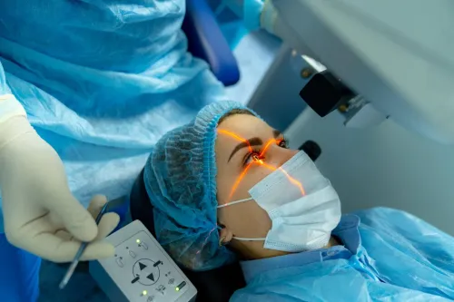
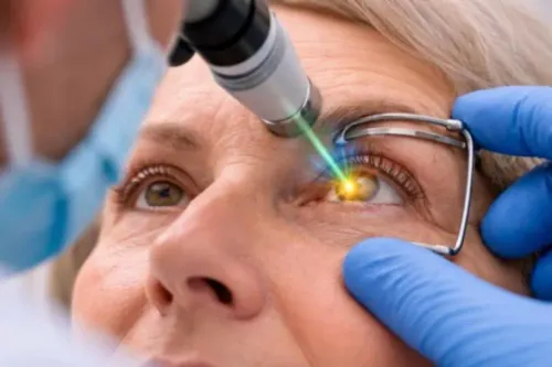
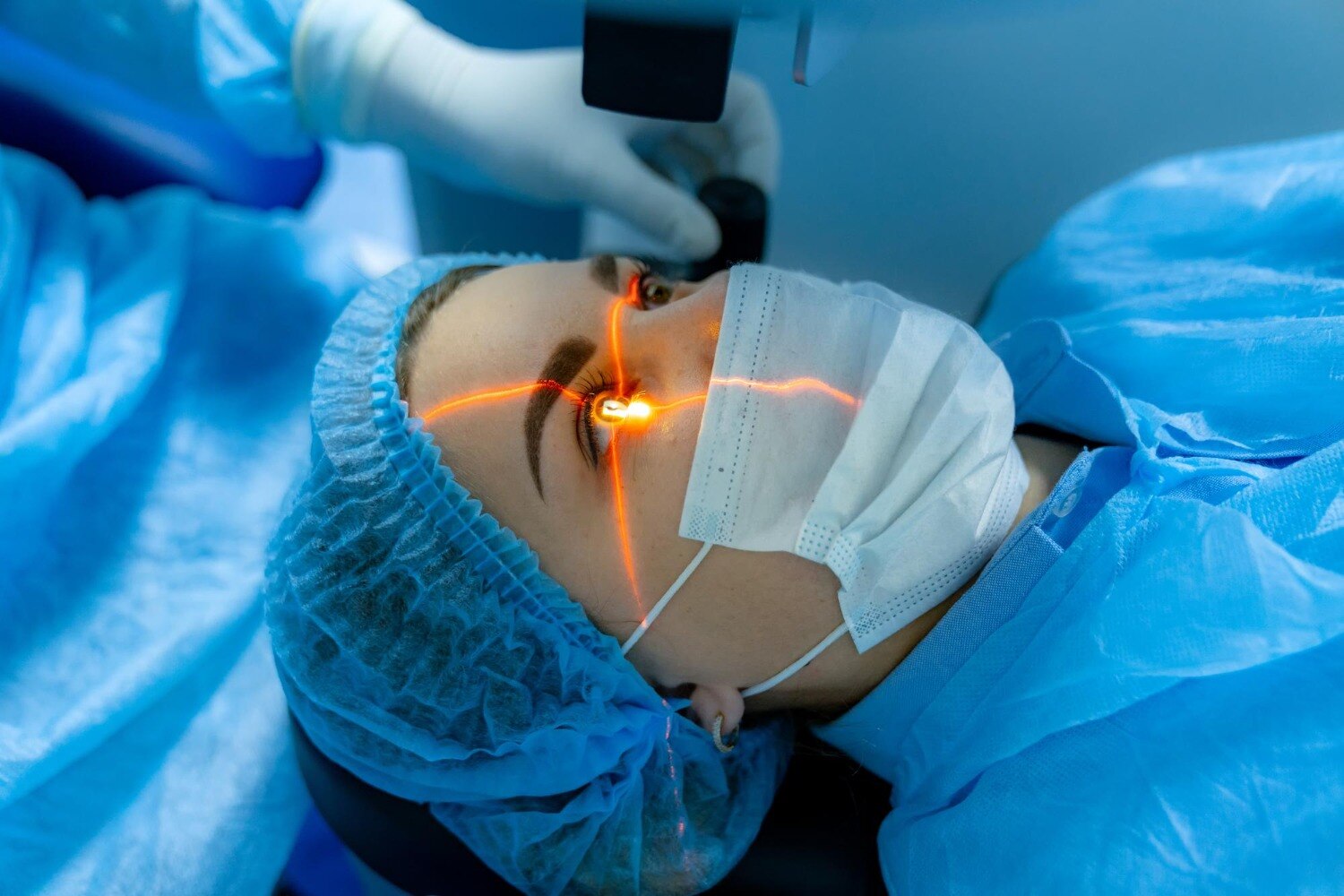
Dilating drops are usually given before retinal laser, followed by a final retinal examination and consent discussion. A local anaesthetic eye laser uses numbing drops; a contact lens keeps the eyelids open and focuses the beam. Expect bright flashes, pressure or brief stinging, but not sharp pain.
Allow about one to two hours for the appointment, including blurred vision after treatment. Arrange transport: driving after laser eye treatment is unsafe while your pupils remain dilated or vision is hazy, so do not drive home or resume driving until your clinician confirms it is safe.
Laser seals a retinal tear or destroys selected peripheral retina; it does not restore already lost vision. Temporary blur, headache, inflammation, raised eye pressure, or light sensitivity can occur. PRP can reduce night or peripheral vision and sometimes worsen macular swelling.
Seek urgent help for severe pain, marked vision loss, new flashes, or a curtain-like shadow.
The safer choice depends on the finding, location, and expected benefit:
| Option | Why it may be safer | What to expect |
|---|---|---|
| Retinal detachment surgery | Detachment, extensive tears, or traction cannot be sealed reliably with laser | Vitrectomy, buckle, or combined surgery |
| Anti-VEGF injections | Macular oedema or active neovascular AMD needs treatment near central vision | Repeat injections and monitoring |
| Cataract treatment | Clouding comes from a cataract, not the retina | Cataract surgery; YAG treats capsule haze after surgery |
| No treatment | A low-risk hole or stable change has no treatment benefit | Observation, warning symptoms, and scheduled review |
At Dr. Jyoti's Hospital, an eye consultation can help separate a retinal problem from cataract or corneal blur before you commit to laser.
Frequently asked questions
Which eye symptoms need urgent retinal assessment?
Arrange a same-day dilated retinal examination for new flashes, a sudden increase in floaters, a curtain or shadow in vision, or sudden vision loss. These symptoms can indicate a retinal tear, retinal detachment, bleeding, or another sight-threatening condition.
When is retinal laser recommended?
Retinal laser is recommended for selected retinal tears, holes, diabetic retinopathy, retinal vein occlusion, and other areas of abnormal or leaking blood vessels. The decision depends on the retinal examination and the location and extent of disease.
What tests determine whether retinal laser is appropriate?
An ophthalmologist may use dilated fundoscopy, optical coherence tomography, retinal photography, ocular ultrasound, or fluorescein angiography. The test depends on whether the concern involves a tear, fluid, bleeding, detachment, or abnormal blood vessels.
How are glaucoma lasers, YAG treatment, and LASIK different?
Glaucoma laser changes fluid drainage or reduces fluid production to lower eye pressure. YAG capsulotomy creates an opening in a cloudy capsule after cataract surgery. LASIK reshapes the cornea to reduce glasses power; it does not treat retinal disease.
When is another option safer than laser treatment?
Observation, injections, cryotherapy, or retinal surgery may be safer when laser cannot reach the affected area, the retina is detached, bleeding blocks the view, or the condition needs a different treatment. Your ophthalmologist should explain the reason and urgency.