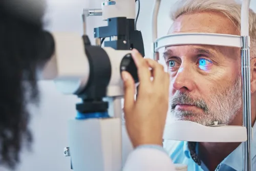
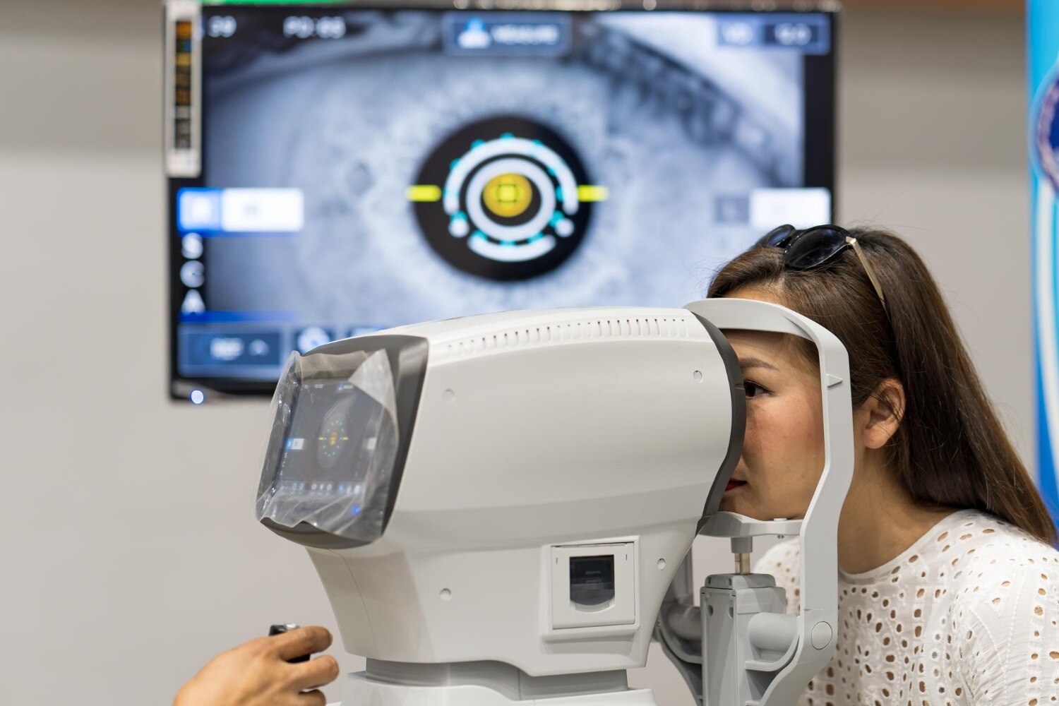
A computerised eye test can estimate how light focuses in your eyes, measure visual performance and produce images or readings that guide further assessment. The meaning depends on which instrument was used, how reliably you responded and whether an eye professional compares the result with a clinical examination and earlier tests.
Key takeaways
- An autorefractor estimates how light focuses; it does not prescribe your final glasses.
- Compare automated readings with subjective refraction and eye-health examinations.
- A clear printout cannot rule out glaucoma, retinal disease, or other eye conditions.
- Seek a comprehensive examination when symptoms, risk factors, or abnormal findings persist.
What an autorefractor measures inside your eyes
An autorefractor estimates how light focuses inside each eye; it does not decide the glasses you should wear. During automated refraction, you look at a target inside the instrument while it analyses reflected light from your eye. Computerised eye testing may also record pupil distance or estimate corneal curvature, depending on the device.
The main refractive results are:
- Sphere power: the strength needed to correct short-sightedness or long-sightedness.
- Cylinder power: the amount of astigmatism detected.
- Cylinder axis: the direction of that astigmatism, recorded in degrees from 1 to 180.
These figures describe the eye’s optical error at that moment. They do not measure eye health, prove that both eyes work together comfortably, or show which lenses you prefer.
The printout is not yet a glasses prescription because the measurement can change when your focusing muscles tighten. Poor fixation, dry eye, a cataract or an irregular corneal shape can also distort the reading. A machine may produce a precise-looking number that does not give you clear, comfortable vision.
An examiner therefore performs subjective refraction after the automated reading. They place trial lenses in front of your eyes and ask which option looks clearer, refining sphere power, cylinder and axis while checking visual clarity and comfort. Your final prescription can differ from the autorefractor result.
What the results reveal about focusing and seeing
Computerised results can show whether the eye focuses light clearly or needs optical correction. Common patterns include:
| Pattern | What the result indicates | What you may notice |
|---|---|---|
| Short-sightedness | The eye focuses distant images in front of the retina | Near objects look clearer than road signs |
| Long-sightedness | The eye focuses images behind the retina | Close work may cause blur or eyestrain |
| Astigmatism | Unequal corneal or lens curvature distorts focus | Blurred or stretched detail at different distances |
| Cylinder power and axis of astigmatism | Cylinder power measures the amount of astigmatic correction; the axis of astigmatism records its orientation from 0 to 180 degrees | The correction may differ between the two eyes |
A visual-acuity test adds a practical measure: it records the smallest high-contrast letter or symbol you can identify at a set distance. Good acuity does not rule out glaucoma, early retinal disease, peripheral-field loss or difficulty seeing in low contrast.
Colour-vision testing can identify difficulty separating colour ranges, commonly along red-green or blue-yellow axes. The result describes test performance; it does not establish whether the cause is inherited, an eye disease, a neurological condition or a medication effect.
An automated visual-field test checks peripheral vision by presenting light points at different locations. Glaucoma, retinal damage and neurological disease can create characteristic loss, but fatigue, poor fixation, learning effects, false-positive responses and false negatives can distort one test. Reliability indices and repeated patterns matter more than an isolated score.
How different instruments examine eye health
These tests answer different questions, so a clear result in one does not replace the others. A routine vision test measures how small high-contrast letters or symbols you identify at a set distance; it does not assess peripheral vision or the optic nerve directly.
| Instrument or test | What it examines | What it can reveal | What it cannot establish |
|---|---|---|---|
| Routine vision test | Central visual acuity | Reduced sharpness, sometimes improved by lenses | Normal acuity does not rule out glaucoma, retinal disease or low-contrast difficulty |
| Visual field test | Peripheral and central field responses | Loss patterns linked to glaucoma, retinal or neurological damage | Fatigue, poor fixation and guessing can distort results |
| Eye-pressure reading | Intraocular pressure (IOP) using computerised tonometry | An unexpectedly high pressure needing confirmation | Normal pressure does not exclude glaucoma; corneal thickness affects readings |
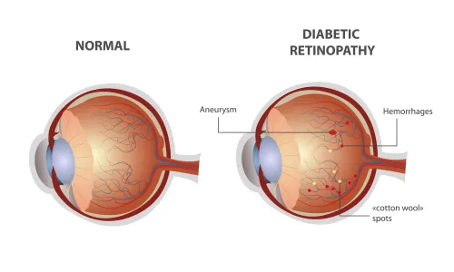
| Retinal image | Photograph of the retina, macula and optic nerve | Visible bleeding, swelling, pigment changes or nerve-head abnormalities | A photograph alone does not confirm every disease |
| Corneal map | Corneal curvature and shape | Irregular astigmatism, ectasia and changes relevant to contact lenses or surgery | It does not measure retinal or optic-nerve health |
| Comprehensive examination | Combined history, vision, refraction, slit-lamp, pressure and retinal assessment | Findings that need comparison, dilation, repeat testing or referral | No single visit guarantees that future disease will not develop |
Automated perimetry is the computerised form of a visual field test: you respond to light flashes at different locations. An examiner interprets its reliability alongside the optic nerve, retinal image and other findings rather than treating any printout as a diagnosis.
Why a normal-looking printout cannot rule out disease
A clean printout shows what the instrument captured, not everything happening inside your eyes. Good visual acuity can coexist with glaucoma, early retinal disease, peripheral-field loss, poor low-contrast vision, or significant cataract severity.
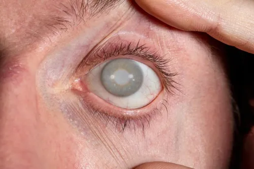
A normal autorefractor reading can also be distorted by dry eye disease, poor fixation, accommodation, an irregular cornea, or a cloudy lens. An air-puff pressure reading does not rule out glaucoma: the disease can occur at statistically normal pressure, and corneal thickness can make the measured value misleading.
Judge the result by checking:
- Reliability indicators: look for poor fixation, excessive blinking, missing responses, false-positive answers, or a low image-quality or signal-strength score. A test marked unreliable cannot support a confident “normal” conclusion.
- Images: a retinal photograph records one moment and may miss the far peripheral retina, subtle changes, or structures hidden by cataract. Compare the image with the examination findings rather than treating it as a verdict.
- Repeatability: when glaucoma, macular disease, or another progressive problem is suspected, repeat testing with comparable settings. A change that appears on one scan but disappears on a well-controlled repeat may reflect measurement noise; a consistent trend deserves assessment.
Symptoms still matter. New flashes, a curtain-like shadow, sudden distortion, a painful red eye, or a rapid drop in vision needs prompt professional evaluation even when the printout looks normal.
When you need more than an automated reading
An automated reading needs professional refinement when it conflicts with your symptoms, changes sharply between attempts, or does not produce consistently clear vision. The examiner performs subjective refraction by comparing lenses, then confirms the cylinder axis, sphere and pupil distance used for spectacles.
Corneal curvature readings also need context because dry eye, contact-lens wear, cataract or an irregular cornea can distort the result.
Ask for a more detailed assessment when:
- You are a child, teenager or young adult with suspected farsightedness, headaches, eyestrain or fluctuating results. Cycloplegic refraction uses drops to temporarily relax focusing; it can reveal the underlying prescription when accommodation is masking hyperopia or creating an over-minus result.
- The prescription seems unexpectedly strong, differs greatly between the eyes, or changes despite careful testing. The professional may repeat the measurement after treating surface dryness or stopping contact lenses for the advised period.
- You have ghosting, double images, distorted lines or poor vision that ordinary lenses do not correct. Corneal topography or tomography can map irregular astigmatism and patterns suggestive of keratoconus.
- You report flashes, new floaters, missing vision, eye pain or sudden blur. Arrange prompt examination rather than relying on refraction.
- A pressure, visual-field or retinal result is abnormal or unreliable. Confirmation can include repeat testing, optic-nerve examination, corneal-thickness measurement, retinal imaging or follow-up over time.
For people considering LASIK, Dr. Jyoti's Hospital can help ensure the final decision uses refined refraction and corneal mapping, not an autorefractor printout alone.
Related service
Eye Care / Ophthalmology Eye Checkup in Zirakpur Get a comprehensive eye checkup in Zirakpur at Dr Jyoti Hospital with experienced eye specialists. View service → |
Frequently asked questions
What does an autorefractor measure?
It estimates how light focuses inside each eye by analysing light reflected from the eye. Depending on the device, it can also record pupil distance or estimate corneal curvature.
Can an autorefractor determine the exact glasses prescription?
No. The reading is a starting estimate. An eye-care professional confirms the prescription with subjective refraction, in which you compare lens choices.
Can a normal computerised eye-test printout rule out eye disease?
No. Automated refraction mainly assesses focusing. It does not replace examinations for conditions such as glaucoma, retinal disease, or corneal problems.
When do you need more than an automated eye reading?
You need further assessment when you have blurred or double vision, eye pain, flashes, new floaters, headaches, unequal vision, or risk factors for eye disease.