
Glaucoma can destroy optic-nerve fibres while your vision still feels normal, and the lost field usually cannot be recovered. You will learn how doctors identify meaningful damage, judge risk beyond one eye-pressure reading, compare treatment choices, and plan follow-up before further loss limits daily life.
Key takeaways
- Glaucoma can permanently damage the optic nerve before symptoms appear.
- A normal eye-pressure reading does not rule out glaucoma.
- Visual fields and optic-nerve imaging reveal damage beyond pressure alone.
- Treatment may include eye drops, laser procedures, or glaucoma surgery.
How Glaucoma Silently Causes Permanent Vision Loss
Glaucoma can damage the optic nerve before you notice pain, blur, or difficulty reading. Open-angle glaucoma usually affects peripheral vision first, leaving central vision intact long enough for you to function normally. This creates peripheral vision loss without symptoms while glaucoma damage continues.
The injury involves optic nerve fibres that carry visual information from the eye to the brain. Once those fibres and their corresponding areas of the visual field are lost, drops, laser treatment, or surgery cannot bring them back.
Existing damage therefore means permanent vision loss; treatment aims to lower eye pressure and prevent or slow further loss.
Early treatment matters because it protects the vision you still have. In the Early Manifest Glaucoma Trial, confirmed progression occurred in about 45% of treated eyes compared with about 62% of untreated eyes during follow-up. Treatment did not guarantee stability, but delaying it surrendered valuable time.
Sudden severe eye pain, a red eye, blurred vision or halos around lights, headache, nausea, or vomiting can signal acute angle-closure glaucoma. Seek emergency eye assessment immediately; this form can cause rapid permanent vision loss.
The Four Glaucoma Categories Doctors Must Distinguish
A normal clinic pressure reading does not rule out glaucoma. Pressure changes during the day, and normal-tension glaucoma damages the optic nerve even when readings stay within a usual range.
| Category | What it means | What distinguishes it |
|---|---|---|
| Open-angle glaucoma | The drainage angle remains open, but fluid leaves too slowly and damages the optic nerve | Gradual, usually painless peripheral-field loss |
| Angle-closure glaucoma | The iris blocks the drainage angle, causing pressure to rise, sometimes suddenly | Severe eye pain, a red eye, halos, headache, nausea, or vomiting require emergency eye care |
| Normal-tension glaucoma | Optic-nerve and visual-field damage occurs without high measured pressure | Lowering pressure still aims to slow progression |
| Glaucoma suspect | Elevated pressure, a suspicious optic nerve, or a thin cornea raises concern, but damage is unconfirmed | Needs surveillance, not an automatic glaucoma diagnosis |
Open-angle and normal-tension disease can progress unnoticed because central vision often remains usable early. Existing optic-nerve fibre loss cannot usually be restored, so the diagnosis determines how urgently pressure-lowering treatment and follow-up are needed.
What Glaucoma Tests Reveal Beyond Eye Pressure
A pressure reading cannot confirm or exclude glaucoma. Goldmann applanation tonometry measures intraocular pressure, but damage can occur at a pressure within the usual reference range.
| Test | What it shows | How to read it |
|---|---|---|
| Goldmann applanation tonometry | Eye pressure in mmHg | A higher number raises risk, but one reading is not a diagnosis; pressure changes during the day. |
| Gonioscopy | Whether the drainage angle is open or closed | The angle determines which glaucoma mechanism is possible and guides treatment. |
| Optic nerve examination | Disc cupping, rim thinning, or an asymmetrical nerve | Compare both eyes and previous photographs; increasing cupping or rim loss is concerning. |
| Optical coherence tomography OCT retinal nerve fibre layer thickness | A cross-sectional image and thickness map of nerve fibres | Thinning, especially on repeat scans, can precede field loss; myopia, unusual discs, or segmentation errors can create false alarms. |
| Automated visual-field testing | Missing or reduced sensitivity, reported in decibels | A repeatable cluster of points and a worsening mean deviation matter more than one missed spot. |
Doctors combine these results because each measures a different part of the disease. A normal pressure does not erase suspicious nerve or field findings, while an abnormal OCT alone does not prove glaucoma.
Poor fixation, fatigue, cataract, or misunderstanding the test can make a visual field unreliable, so repeat testing and comparison with earlier images are essential.
Why Risk and Follow-Up Matter Even With a Normal Pressure Reading
A normal intraocular pressure reading does not guarantee a safe optic nerve. Pressure changes during the day, and normal-tension glaucoma can progress despite ordinary clinic readings; thinner corneas can also make measured pressure underestimate risk.
Risk is higher with:
- Older age or a family history of glaucoma
- High myopia, diabetes, steroid use, or a previous eye injury
- Higher pressure, a larger cup-to-disc ratio, or abnormalities on automated visual-field testing
Follow-up intervals differ because doctors weigh risk, existing damage, and evidence of change rather than pressure alone. OCT scans, optic-nerve examinations, and automated visual-field testing can disagree, so one unreliable test should not determine the schedule.
| Patient profile | Why monitoring changes |
|---|---|
| Lower-risk suspect with normal nerve and reliable tests | Longer intervals may be reasonable if results remain stable. |
| Higher-risk suspect with a large cup-to-disc ratio, thinner corneas, high myopia, or strong family history | Closer review detects structural or field changes earlier. |
| Established damage, worsening OCT, or repeatable field loss | Shorter intervals and a lower target pressure protect remaining vision. |
A missed appointment matters most when damage is advancing silently. Ask how often you need review, which result would shorten that interval, and whether your pressure target reflects the condition of both eyes.
Comparing Drops, Laser Procedures, and Glaucoma Surgery
Treatment choice depends on glaucoma type, target eye pressure, existing damage, and how reliably you can follow a plan. No option restores lost optic-nerve fibers or visual-field loss; each aims to prevent further damage. Compare the trade-offs with your ophthalmologist.
| Option | Main benefit | Limitations, side effects, and long-term demands |
|---|---|---|
| Prescription pressure-lowering eye drops | Lower pressure without a procedure; useful across many glaucoma types | Require regular dosing indefinitely. Burning, redness, blurred vision, difficulty instilling drops, cost, and missed doses can leave pressure above target. |
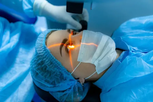
| Selective laser trabeculoplasty | Treats open-angle glaucoma and can work as a first treatment, reducing dependence on drops | Not suitable for every angle or disease stage. Pressure can rise briefly; effect may fade, and repeat treatment or drops may become necessary. Most people resume normal activity quickly. |
| Laser peripheral iridotomy | Creates a tiny opening to relieve or prevent angle closure | Designed for angle-closure risk, not routine open-angle glaucoma. Expect light sensitivity or inflammation for several days; it does not remove established damage. |
| Minimally invasive glaucoma surgery MIGS | Modest pressure reduction with less tissue disruption, often during cataract surgery | May not reach a low enough target alone. Expect short recovery, but continued drops and monitoring remain possible. |
| Trabeculectomy or glaucoma drainage devices | Stronger pressure lowering for advanced or uncontrolled disease | Higher risks include infection, bleeding, pressure that is too low, and cataract. Recovery and medication adjustments take weeks; follow-up remains intensive for life. |
Choosing the least burdensome effective option matters, but delaying treatment risks irreversible progression.
Building a Safe Long-Term Glaucoma Plan
Expect the visit to review your eye pressure, optic nerve, OCT scans, visual-field results, medicines, side effects, and target pressure. Confirm the glaucoma consultation duration and whether pupil dilation blurred vision will make driving unsafe afterward. Dr. Jyoti's Hospital can help you organise a baseline eye record when previous reports are scattered across clinics.
Bring these details:
- Every eye drop, dose, bottle, allergy, and supplement
- Previous pressure readings, scans, visual fields, and operation notes
- A written record of missed doses, irritation, difficulty using drops, and refill problems
- Questions about punctal occlusion, missed doses, refill planning, and whether preservative-free drops are suitable
- A direct comparison of cataract surgery versus glaucoma treatment if cloudy vision and pressure control are both concerns
Routine follow-up is not enough for sudden severe eye pain, a red eye, halos, blurred vision, headache, nausea, or vomiting. Seek immediate emergency eye assessment; acute angle closure can permanently damage vision quickly. Also contact your eye specialist sooner if vision changes, treatment is intolerable, or pressure remains above target despite taking drops correctly.
Frequently asked questions
How does glaucoma cause permanent vision loss?
Glaucoma damages the optic nerve, often starting with peripheral vision while central vision remains clear. Untreated damage is permanent.
What tests detect glaucoma beyond eye pressure?
Doctors assess the optic nerve, measure visual fields, and use retinal or optic-nerve imaging alongside eye-pressure readings.
Can you have glaucoma with normal eye pressure?
Yes. Normal-tension glaucoma causes optic-nerve damage despite pressure readings within the usual range, so risk assessment and follow-up remain important.
How is glaucoma treated?
Treatment may use pressure-lowering eye drops, laser procedures, or glaucoma surgery, depending on the glaucoma category, damage, and response to treatment.