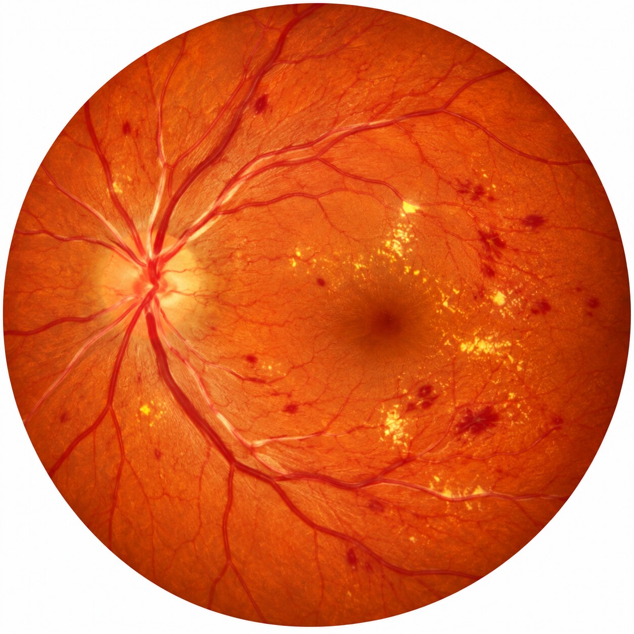
High blood pressure can remodel retinal arterioles, disrupt the blood-retina barrier, and injure the macula or optic disc. You will learn what these changes look like, which symptoms require urgent care, how specialists distinguish chronic vessel damage from an acute hypertensive emergency, and which tests clarify the cause.
Key takeaways
- Persistent high blood pressure narrows and thickens retinal arterioles.
- Retinal bleeding, swelling, and cotton-wool spots can signal vascular damage.
- Retinopathy, choroidopathy, and optic neuropathy affect different eye structures.
- Sudden vision loss, severe headache, or confusion needs emergency assessment.
How High Blood Pressure Changes the Retinal Blood Vessels
Persistently high blood pressure forces retinal arterioles to constrict and remodel. Their walls become thickened, making the inner channel narrower; this arteriolar narrowing reduces blood flow to the retina and increases resistance within the vessel. These chronic changes are central to hypertensive retinopathy.
At arteriovenous crossings, a thickened arteriole can press on the vein beneath it. The venous column then appears interrupted or tapered, a sign called arteriovenous nicking. It indicates chronic vascular remodeling, not proof of a current hypertensive crisis.
If pressure rises severely or abruptly, the retinal blood-retina barrier can fail. Fluid and blood escape into surrounding tissue, while damaged small vessels stop supplying parts of the retina. The examination may show:
- Retinal edema from leaked fluid
- Flame-shaped or dot-and-blot haemorrhages from ruptured vessels
- Cotton-wool spots caused by small areas of nerve-fibre-layer ischemia
- Hard exudates made from deposited blood lipids
- Optic-disc swelling, which signals severe injury and needs urgent medical assessment
Long-standing high pressure can leave permanent narrowing and ischemic damage even after treatment. Controlling blood pressure can reduce leakage, haemorrhages, and swelling, but no eye drop reverses established vessel-wall thickening or arteriovenous nicking.
What Retinal Lesions Mean for Vision and Overall Health
Retinal lesions reduce vision mainly when they involve the macula, optic nerve, or retinal circulation. Damage farther from the centre can be extensive while reading and face recognition remain normal.
| Lesion | Effect on vision | What it indicates |
|---|---|---|
| Retinal haemorrhages | Blur or blind spots when near the macula | Leaking or ruptured retinal vessels |
| Cotton-wool spots | Usually no immediate symptom; a larger area of ischemia can affect vision | Localised interruption of nerve-fibre blood flow |
| Hard exudates | Reduced central vision when deposits reach the macula | Leakage of serum or lipids |
| Macular star or macular edema | Distorted, blurred, or reduced central vision | Macular leakage and swelling |
| Optic-disc swelling | Blurred vision, enlarged blind spot, or rapid loss of sight | Possible severe pressure-related injury requiring urgent assessment |
A person can have substantial damage without noticing it because slow changes allow the visual system to adapt, and lesions outside the macula do not affect detailed central sight. The retina also has no pain sensation, so tissue injury does not announce itself like an eye infection or surface injury.
Retinal haemorrhages, cotton-wool spots, and hard exudates show vascular damage, but they do not reveal the blood-pressure reading at that moment. Their pattern can signal broader vessel injury involving organs such as the kidneys, brain, or heart, so clinicians interpret the eye findings alongside blood-pressure measurement and other symptoms.
Sudden blur, field loss, or vision loss needs urgent medical assessment.
Retinopathy, Choroidopathy, and Optic Neuropathy Are Not the Same
A sudden, severe rise in blood pressure can injure three separate eye structures: the retinal vessels, the choroid beneath the retina, and the optic nerve. The resulting findings overlap, but they do not mean the same thing.
| Condition | Main site | What severe pressure can do |
|---|---|---|
| Hypertensive retinopathy | Retinal arterioles and retinal tissue | Narrowed or thickened vessels leak fluid, causing hemorrhages, cotton-wool spots, hard exudates, or retinal edema. |
| Hypertensive choroidopathy | Choroidal arterioles and retinal pigment epithelium | Ischemic injury can produce Elschnig spots, which are pale or dark patches, and Siegrist streaks, which are linear pigment changes along choroidal vessels. |
| Hypertensive optic neuropathy | Optic nerve and optic disc | Reduced blood flow and swelling can impair nerve function, causing blurred vision, blind spots, or visual-field loss. |
Hypertensive choroidopathy reflects damage below the retina, so its signs are not simply advanced retinal-vessel narrowing or ordinary age-related spots. It is more likely with abruptly accelerated or extremely high pressure.
Swelling of the optic disc is the most urgent distinction. Hypertensive optic neuropathy with disc edema suggests a hypertensive emergency and possible injury to the brain, kidneys, heart, or other organs; it requires immediate medical assessment, not a routine eye appointment.
A clinician must also separate it from papilledema caused by raised pressure inside the skull.
How an Eye Specialist Stages and Investigates the Damage
A specialist begins by checking visual acuity, pupils, eye pressure, and the symptoms’ timing, then reviews blood-pressure readings, treatment, diabetes, kidney disease, and smoking history. These details help separate chronic vascular change from a sudden pressure-related injury or another retinal disorder.
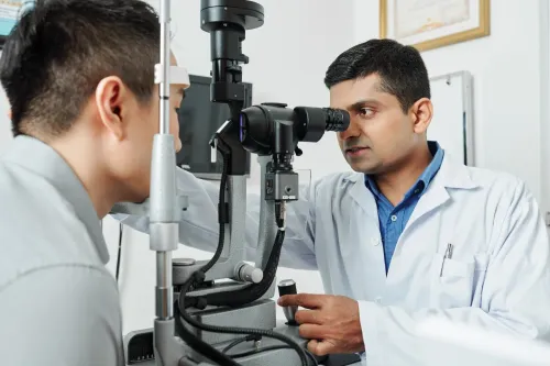
The key examination is a dilated fundus examination. After drops widen the pupils, the clinician inspects the retinal arterioles, crossings, macula, optic disc, and surrounding retina for narrowing, vessel-wall changes, haemorrhages, cotton-wool spots, hard exudates, leakage, or disc swelling.
The Keith-Wagener-Barker grading system provides a historical framework:
- Grade 1: mild, generalised arteriolar narrowing.
- Grade 2: narrowing with arteriovenous nicking and more obvious vessel-wall change.
- Grade 3: Grade 2 findings plus haemorrhages, cotton-wool spots, or hard exudates.
- Grade 4: Grade 3 findings plus optic-disc swelling.
Clinicians do not use this scale alone. Examiner disagreement is possible, and isolated arteriolar narrowing does not prove the current blood pressure or predict cardiovascular risk by itself. Fundus photography records findings for comparison, while optical coherence tomography shows macular swelling or retinal-layer injury.
Fluorescein angiography is reserved for selected cases involving suspected leakage or ischaemia.
At Dr. Jyoti's Hospital, ask whether imaging is needed when photographs or the dilated examination show macular or optic-disc changes. A sudden visual-field defect or loss of vision needs urgent assessment, not a routine follow-up.
Related service
Eye Care / Ophthalmology Eye Checkup in Zirakpur Get a comprehensive eye checkup in Zirakpur at Dr Jyoti Hospital with experienced eye specialists. View service → |
When Retinal Findings Mean You Need Emergency Assessment
A blood-pressure reading of 180/120 mm Hg or higher, combined with sudden vision change, severe headache, confusion, weakness, chest pain, or shortness of breath, requires immediate medical assessment. Do not wait for a routine eye appointment.
Optic-disc swelling with markedly elevated pressure is also an emergency warning because it can indicate a hypertensive emergency affecting the brain, kidneys, heart, or eyes.
Seek emergency help immediately for:
- New blurred or lost vision, a missing area of vision, or sudden flashes and floaters
- Severe headache, confusion, trouble speaking, weakness, or numbness
- Chest pain, breathing difficulty, or fainting
Retinal damage can exist without symptoms, so a high reading still needs prompt medical review even when your sight feels normal. Doctors may repeat the pressure, examine the optic nerve and retina, and use fluorescein angiography to identify leakage or poor retinal blood flow; testing must not delay emergency treatment.
| Urgency | Typical situation | What happens next |
|---|---|---|
| Routine review | Retinal narrowing without severe pressure or symptoms | Confirm blood-pressure history and arrange specialist follow-up |
| Urgent assessment | Sudden vision change or new retinal hemorrhages | Examine the eye and investigate the cause promptly |
| Emergency care | Pressure at least 180/120 mm Hg plus symptoms or optic-disc swelling | Monitor organs and lower pressure carefully; reducing it too fast can also cause damage |
Frequently asked questions
How does high blood pressure change retinal blood vessels?
Persistent hypertension causes retinal arterioles to constrict and remodel. Their walls thicken, the inner channel narrows, and blood-flow resistance rises.
What can retinal lesions mean for vision and health?
Lesions such as hemorrhages, retinal swelling, and cotton-wool spots indicate damage from raised blood pressure. They can threaten vision and signal wider vascular injury.
Are hypertensive retinopathy, choroidopathy, and optic neuropathy the same?
No. Retinopathy affects retinal vessels and tissue, choroidopathy affects the choroid beneath the retina, and optic neuropathy affects the optic nerve.
How does an eye specialist investigate hypertensive retinal damage?
The specialist examines the retina after pupil dilation and may use retinal photography, optical coherence tomography, or fluorescein angiography. Blood-pressure measurement and medical assessment help identify the cause and severity.
When do retinal findings require emergency assessment?
Seek emergency care for sudden vision loss, severe headache, confusion, weakness, chest pain, breathlessness, or very high blood pressure with visual symptoms.