A green retinal laser does not cut out damaged tissue or restore vision that disease has already destroyed. It delivers carefully controlled heat to selected retinal areas, and the treatment plan depends on whether the problem is leakage, a retinal break, oxygen-starved tissue, or abnormal blood-vessel growth.
Key takeaways
- Green laser creates controlled retinal burns using absorbed light and heat.
- Your ophthalmologist checks retinal findings, leakage, bleeding, and treatment goals first.
- Focal, grid, and panretinal patterns target different retinal problems.
- Injections or surgery may fit better when disease affects the macula or vitreous.
What Green Laser Does in Retinal Care
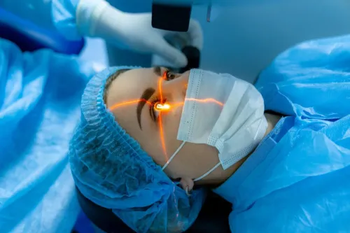
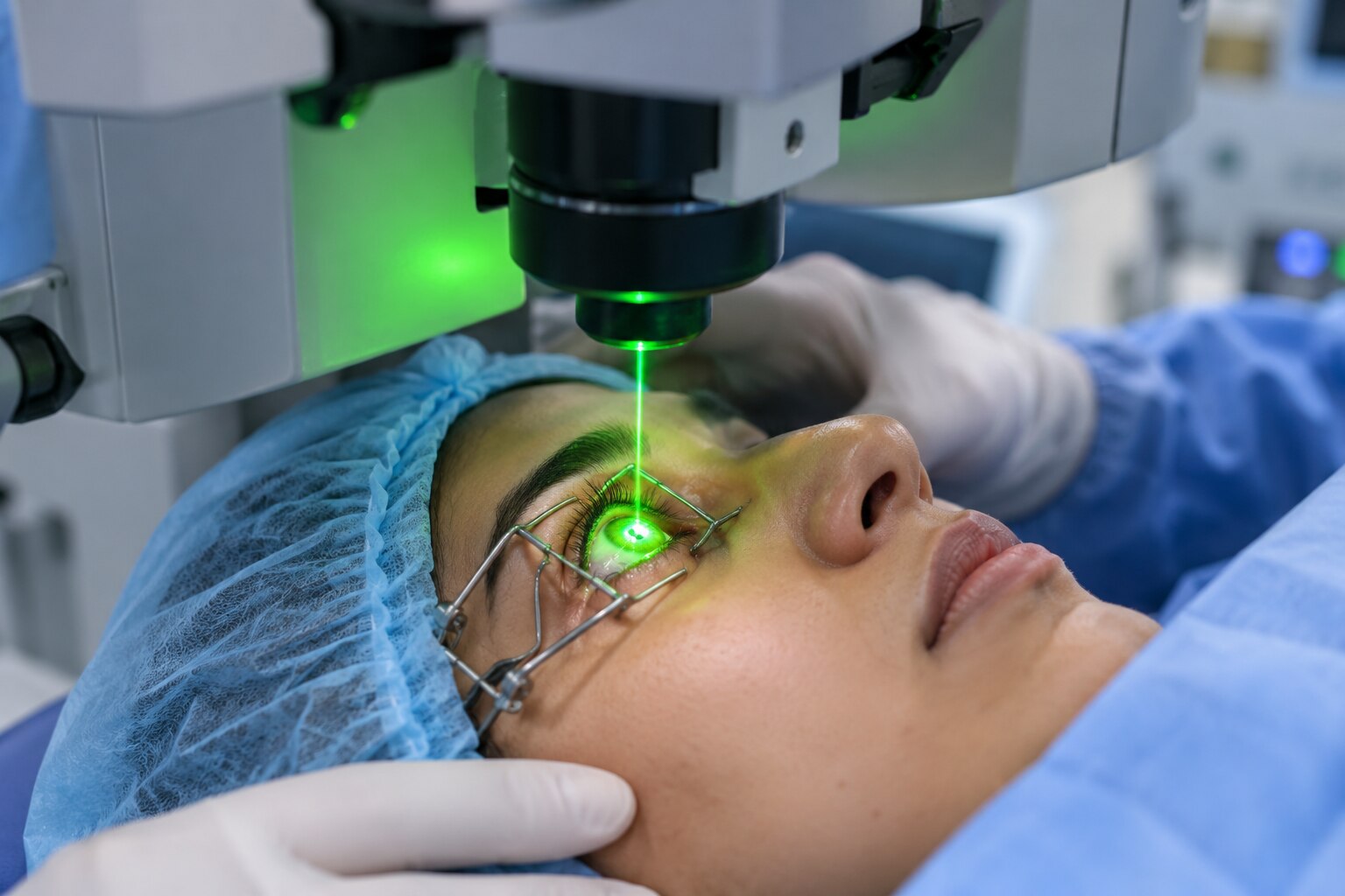
A green laser in retinal care usually means a 532-nanometre laser, produced by an argon or frequency-doubled green laser system. It is used for retinal photocoagulation, also called laser photocoagulation. Melanin and haemoglobin absorb the light and convert it into heat, creating controlled thermal burns in the retinal pigment epithelium and choroid.
The laser heats tissue; it does not cut or vaporise the retina.
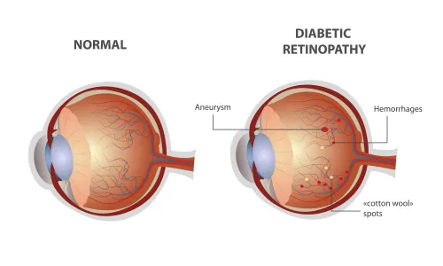
- Proliferative diabetic retinopathy: treats oxygen-starved peripheral retina, reducing the signal that drives abnormal blood-vessel growth.
- Selected diabetic macular oedema: targets leaking areas to reduce fluid leakage.
- Retinal tears or holes: places burns around the break, forming a scar that helps seal its edge.
- Lattice degeneration with risk features: reinforces vulnerable retinal areas to reduce the risk of a future tear or detachment.
- Retinal vein occlusion with abnormal new vessels: treats ischaemic retina and limits further abnormal vessel growth.
- Retinal neovascularisation: limits the spread and activity of fragile new vessels that can bleed.
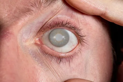
Laser cannot restore retina already destroyed by macular disease, bleeding, optic-nerve damage, or retinal detachment. Its purpose is to protect remaining vision or prevent a retinal problem from worsening; it cannot reverse established structural loss.
How the Ophthalmologist Decides Whether Laser Is Suitable
Laser suitability starts with the retinal finding, not the fact that the light is green. A dilated retinal examination lets the ophthalmologist inspect bleeding, abnormal new vessels, retinal detachment risk, and the location of a tear or hole. Fundus photography records the appearance for comparison during follow-up.
Optical coherence tomography measures retinal thickness and shows whether swelling involves the macula or fovea. Fluorescein angiography is added when the blood-vessel pattern or leakage needs clarification, helping identify leaking areas or ischaemic retina that cannot be judged reliably from examination alone.
The clinician also checks whether laser can reach and focus on the target. Cataract, vitreous haemorrhage, another media opacity, poor fixation, or dense retinal swelling can block the view or make accurate delivery impossible. Before treatment, the specialist considers:
- Retinal pigmentation, tissue thickness, media clarity, and distance from the fovea when setting power and focus
- A mild gray-white retinal reaction as the usual endpoint, rather than an intense white burn
- Excessive power or poor focus, which can cause foveal injury, haemorrhage, choroidal damage, or an enlarging scar
A suitable plan might therefore use laser, another wavelength, injection, cryotherapy, or surgery. The examination determines that choice.
What Happens During and After Retinal Laser Treatment
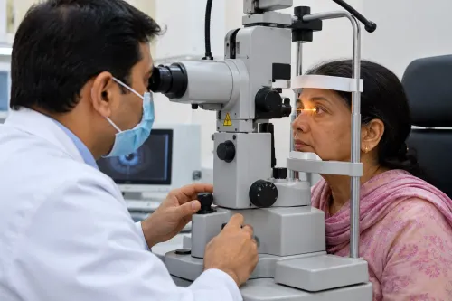
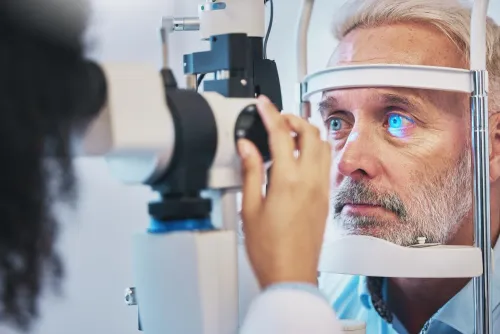
The appointment usually takes place with you seated at a slit lamp. Numbing eye drops are given, then a contact lens is placed on the eye to steady it and focus the ophthalmologist’s view of the retina. Through the slit lamp, the ophthalmologist delivers precise laser burns.
You may notice:
- Bright flashes with each group of laser spots
- Pressure from the contact lens
- Discomfort or a brief aching sensation
- Temporary blurred vision
- Light sensitivity after the procedure
The number and placement of spots depend on the retinal condition and the treatment goal. For a retinal tear or hole, laser retinopexy places burns around the break. A chorioretinal scar develops over about one to two weeks and helps seal its edge.
Laser retinopexy does not remove existing floaters or repair a retina that has already detached. Arrange transport if your vision is affected, and do not drive until your vision is clear and the treating clinician permits it.
Seek an immediate retinal examination if you develop new flashes, a sudden increase in floaters, or a curtain-like defect in your field of vision. These symptoms can signal a new tear elsewhere, even after successful treatment of the original break.
Focal, Grid, and Panretinal Laser: How the Treatment Pattern Changes
Laser spots follow the retinal problem, because a leaking point, swollen macula, and oxygen-starved peripheral retina need different treatment patterns.
- Focal laser targets specific leaking microaneurysms or other abnormal areas. The burns are placed where leakage originates rather than spread across healthy retina.
- Grid laser applies a planned pattern over retinal thickening or leakage, usually away from the fovea and other delicate central structures. It treats a zone, not one identifiable leaking vessel.
- Panretinal photocoagulation, or PRP, treats broad areas of peripheral ischaemic retina in proliferative diabetic retinopathy. It reduces the stimulus for abnormal vessels growing on the retina or optic disc; it is not aimed only at visible new vessels.
PRP mainly reduces the risk of severe future visual loss. It cannot restore vision already lost from macular damage, haemorrhage, detachment, or optic-nerve disease, and more than one session may be needed when new vessels persist or recur.
The trade-off is reduced peripheral or night vision, a smaller visual field, and lower contrast sensitivity. PRP can also worsen or trigger diabetic macular oedema, so the macula must be assessed and treated separately.
For centre-involving diabetic macular oedema with reduced vision, anti-VEGF injections are generally preferred. Focal or grid laser retains a selective role for non-centre-involving oedema or persistent leakage.
Retinal vein occlusion with macular oedema is commonly treated first with anti-VEGF medication or corticosteroid. Grid laser is reserved for selected leakage patterns, not every case.
When Laser Is the Right Tool—and When Another Retinal Treatment Fits Better
“Green” does not decide treatment; the retinal finding does. A macula-threatening retinal detachment, tractional retinal detachment, or dense vitreous haemorrhage blocking the laser view can require vitrectomy or other urgent retinal surgery instead.
| Option | Main trade-off | When it fits |
|---|---|---|
| Observation | Avoids treatment risk, but requires surveillance | Low-risk retinal findings without active leakage, dangerous new vessels, or detachment |
| Laser photocoagulation | Seals or suppresses disease, but can scar functioning retina and reduce peripheral or night vision | A visible, reachable tear, leaking focus, or peripheral ischaemic retina |
| Cryotherapy | Treats through tissue when laser access is poor, but causes more inflammation and less precise scarring | A retinal break that cannot be reached effectively with laser |
| Anti-VEGF injections | Improves leakage or abnormal vessel activity, but requires repeat treatment and close follow-up | Appropriate macular oedema, diabetic macular oedema, or retinal neovascularisation |
Anti-VEGF injections can also replace or supplement panretinal photocoagulation (PRP) in proliferative diabetic retinopathy. In the DRCR.net Protocol S trial, ranibizumab produced visual acuity noninferior to PRP at two years, with less visual-field loss and less diabetic macular oedema; repeated injections and close follow-up remained necessary.
Another wavelength may be chosen when cataract, haemorrhage, or another media opacity prevents green light from reaching the target, or when the tissue does not absorb it adequately. In Zirakpur, a specialist retinal assessment at Dr. Jyoti's Hospital can match the examination findings to laser, injections, surgery, cryotherapy, or observation.
Frequently asked questions
What does a green laser do in retinal care?
A 532-nanometre green laser delivers focused light absorbed by melanin and haemoglobin. The absorbed energy creates controlled thermal burns for retinal photocoagulation.
How does an ophthalmologist decide whether retinal laser is suitable?
The ophthalmologist examines the retina and considers the condition, location of abnormal vessels or leakage, vision risk, and the treatment goal before selecting laser.
What happens during and after retinal laser treatment?
You receive anaesthetic drops or another form of eye anaesthesia, then look at a target while the ophthalmologist applies laser spots. Blurred vision, light sensitivity, or discomfort can follow, so arrange transport and follow the clinic’s aftercare instructions.
What is the difference between focal, grid, and panretinal laser?
Focal laser treats selected leaking points, grid laser places a wider pattern over an area of retinal swelling, and panretinal laser treats larger peripheral retinal areas affected by abnormal blood-vessel growth.
When might another retinal treatment be better than green laser?
Anti-VEGF injections, corticosteroid treatment, cryotherapy, or vitrectomy may fit better when the disease involves the macula, vitreous haemorrhage, traction, or another problem that laser cannot adequately treat.